
A magnetic resonance imaging (MRI) scan uses a strong magnetic field and radio waves to create detailed images of the organs and tissues within the body. MRI can help with viewing injuries, tumors, certain heart problems, and more.
Since its invention, doctors and researchers continue to refine MRI techniques to assist in medical procedures and research. The development of MRI revolutionized medicine.
This article looks specifically at MRI scans, how they work, and how doctors use them.
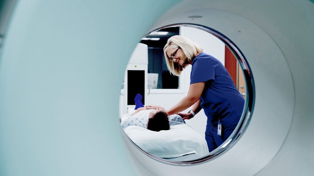
An MRI scan uses a large magnet, radio waves, and a computer to create a detailed, cross-sectional image of internal organs and structures. The scanner itself typically resembles a large tube with a table in the middle, allowing the patient to slide in.
An MRI scan can examine many parts of the body, including:
- brain and spinal cord
- joints and bones
- breasts
- blood vessels
- the heart
- internal organs, such as the prostate, womb, or liver
An MRI scan differs from CT scans and X-rays, as it
Functional magnetic resonance imaging (fMRI)
Functional magnetic resonance imaging, or functional MRI (fMRI), uses MRI technology to measure cognitive activity by monitoring blood flow to certain areas of the brain.
Doctors use fMRI to:
- asses which part of the brain is handling functions such as thought, speech sensation, and movement, called brain mapping
- assess the growth of brain tumors
- help plan surgery, radiation therapy, or other invasive treatments for the brain
The following are examples in which an MRI scanner would be used:
- anomalies of the brain and spinal cord
- tumors, cysts, and other anomalies in various parts of the body
- breast cancer screening for those with a high risk of developing the condition
- injuries or abnormalities of the joints, such as the back and knee
- certain types of heart problems
- diseases of the liver and other abdominal organs
- the evaluation of pelvic pain, with causes including fibroids and endometriosis
- suspected uterine anomalies in those undergoing evaluation for infertility
This list is by no means exhaustive. The use of MRI technology is always expanding in scope and use.
There is very little preparation required, if any, before an MRI scan.
On arrival at the hospital, doctors may ask the patient to change into a gown. If a gown is not required, a person can wear clothes that do not contain any metal, such as zips, belts, buckles, or bras with fasteners or underwires.
This is because an MRI machine uses magnets. It is also important to remove any other metal objects, such as:
- jewellery
- watches
- piercings
- dentures
- hearing aids
- wigs
Individuals who are anxious or nervous about enclosed spaces should tell the doctor. A doctor may be able to prescribe medication to help ease the anxiety and make the procedure more comfortable.
Patients will sometimes receive an injection of intravenous (IV) contrast liquid to improve the visibility of a particular tissue that is relevant to the scan.
The radiologist, a doctor who specializes in medical images, will then talk the individual through the MRI scanning process and answer any questions they may have about the procedure.
Children and babies may need to undergo general anesthesia, as it is important to remain perfectly still during the procedure.
During the scan, a person can have a friend or family member stay with them. The friend or family member will also need to remove any metal objects and follow the same clothing guidelines.
Once the person has entered the scanning room, the doctor will help them onto the scanner table to lie down. Staff will ensure that they are as comfortable as possible by providing blankets or cushions.
Earplugs or headphones will be provided to block out the loud noises of the scanner. The latter is popular with children, as they can listen to music to calm any anxiety during the procedure.
Once in the scanner, the MRI technician communicates with the person via the intercom to ensure they are as comfortable as possible. They will not start the scan until the person is ready.
During the scan, it is vital to stay still. Any movement will disrupt the images, much like a camera trying to take a picture of a moving object. The scanner will make loud clanging noises, which is perfectly normal.
Depending on the images, at times it may be necessary for the person to hold their breath.
If the person feels uncomfortable during the procedure, they can speak to the MRI technician via the intercom and request that the scan be stopped.
After the scan, the radiologist examines the images to determine whether any more are required. If the radiologist is satisfied, the patient can go home.
The radiologist will prepare a report for the requesting doctor. Patients are usually asked to make an appointment with their doctor to discuss the results.
MRIs are one of the safest medical procedures available.
However, the contrast dye can cause nausea, headaches, and pain or burning at the point of injection in some people.
Allergy to the contrast material is also possible, and it can cause hives or itchy eyes. If any adverse reactions occur, the person should notify the technician.
People who experience claustrophobia or feel uncomfortable in enclosed spaces sometimes express difficulties with undergoing an MRI scan.
An MRI scanner contains powerful magnets. These magnets
When the radio waves stop, the protons shift back into place and release energy. The MRI machine detects how much and how quickly this energy is released, depending on the type of tissue. Radiologists can tell these types of tissue apart.
Although the person cannot feel these changes, the scanner can detect them and, in conjunction with a computer, can create a detailed cross-sectional image for the radiologist. If a dye is injected, it can result in brighter and clearer images because it helps the protons to realign more quickly.
The United Kingdom’s National Health Service (NHS) states that a single scan can take a few minutes, up to 3 or 4 minutes, and the entire procedure can take 15 to 90 minutes.
Typically, a person will be able to eat, drink, or take any medication unless a healthcare professional instructs otherwise. However, depending on the part of the body being examined, a person may not be able to eat or drink up to 4 hours before the scan. Alternatively, a person may need to drink a large amount of water before the scan.
Although braces and fillings are unaffected by the scan, they may distort certain images. The doctor and technician will discuss this beforehand. The MRI scan may take longer if additional images are required.
It is important to stay as still as possible while in the MRI scanner. Any movement will distort the scanner, producing blurry images. In particularly long MRI scans, the MRI technician may allow a short break halfway through the procedure.
The doctor and radiologist will be able to talk the person through the whole procedure and address any anxieties. To help those with claustrophobia, open MRI scanners are available in some locations for certain body parts.
A person can take medication prior to the test to ease anxiety.
A contrast dye can improve diagnostic accuracy by highlighting certain tissues. Some people may need to have a contrast agent injected before the scan.
A person should let the healthcare professional know about the pregnancy before an MRI.
According to guidelines suggested by the American College of Obstetricians and Gynecologists, MRIs are not associated with risk and are a good imaging choice for pregnant people. However, healthcare professionals should only use them when it is necessary and provides medical benefit to the pregnant person.
Using gadolinium contrast dye should only be used if it significantly improves the diagnostic performance, or improves the outcome for the fetus or pregnant person.